45 structure of the heart with labels
Human Heart - Anatomy, Functions and Facts about Heart - BYJUS The external structure of the heart has many blood vessels that form a network, with other major vessels emerging from within the structure. The blood vessels typically comprise the following: Veins supply deoxygenated blood to the heart via inferior and superior vena cava, and it eventually drains into the right atrium. Heart: Anatomy and Function The parts of your heart are like the parts of a house. Your heart has: Walls. Chambers (rooms). Valves (doors). Blood vessels (plumbing). Electrical conduction system (electricity). Heart walls Your heart walls are the muscles that contract (squeeze) and relax to send blood throughout your body.
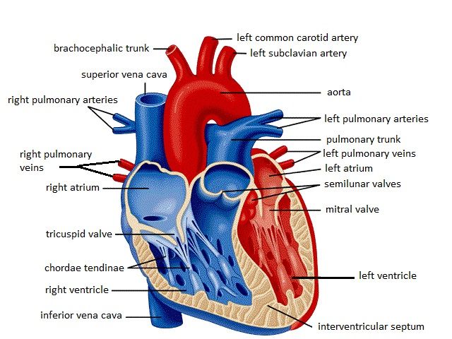
Label Heart Anatomy Diagram Printout - Enchanted Learning Every day, the heart pumps about 2,000 gallons (7,600 liters) of blood, beating about 100,000 times. Label the heart anatomy diagram below using the heart glossary . Note: On the diagram, the right side of the heart appears on the left side of the picture (and vice versa) because you are looking at the heart from the front.

Structure of the heart with labels
PDF Anatomy of Heart Labeled and Unlabeled Images (a) Anterior view of the external heart C' 2019 Pearson Education. Aort'c arch Ligamentum arteriosum Left pulmonary artery Left pulmonary ve ns Auricle of left atrium Circumflex artery Left coronary artery (in atrioventricular sulcus) Great cardiac vein Left ventricle Anterior interventricular artery (in anterior interventricular sulcus) Apex Heart Anatomy | Anatomy and Physiology - Lumen Learning Internal Structure of the Heart. Recall that the heart's contraction cycle follows a dual pattern of circulation—the pulmonary and systemic circuits—because of the pairs of chambers that pump blood into the circulation. In order to develop a more precise understanding of cardiac function, it is first necessary to explore the internal ... Heart Blood Flow | Simple Anatomy Diagram, Cardiac ... The anatomy of the heart was made easy in a previous EZmed video and post, where we learned tricks to remember the main cardiac structures shown below. Check out the anatomy of the heart linked below, as that will be a great review of the main cardiac structures before learning the blood flow! Heart Anatomy: Labeled Diagram, Structures ...
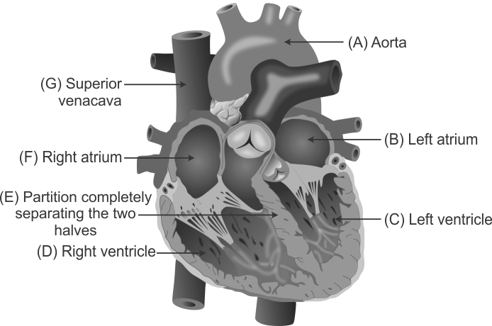
Structure of the heart with labels. Lesson | The Heart - External Structure | Encounter Edu In this lesson students begin their exploration of the circulatory system, labelling a diagram of the external structures and identifying arteries and veins. Heart Anatomy With Labels Photos and Premium High Res ... Browse 147 heart anatomy with labels stock photos and images available, or start a new search to explore more stock photos and images. heart blood flow - heart anatomy with labels stock illustrations. human heart anatomy. blood flow - heart anatomy with labels stock illustrations. circulatory system, diagram - heart anatomy with labels stock ... Labelling the heart — Science Learning Hub Blood transports oxygen and nutrients to the body. It is also involved in the removal of metabolic wastes. In this interactive, you can label parts of the human heart. Drag and drop the text labels onto the boxes next to the diagram. Selecting or hovering over a box will highlight each area in the diagram. The Anatomy of the Heart, Its Structures, and Functions The heart is the organ that helps supply blood and oxygen to all parts of the body. It is divided by a partition (or septum) into two halves. The halves are, in turn, divided into four chambers. The heart is situated within the chest cavity and surrounded by a fluid-filled sac called the pericardium. This amazing muscle produces electrical ...
Structure and Function of the Heart - News-Medical The heart also has a wall that is composed of three layers: the outer layer epicardium (thin layer), the middle layer myocardium (thick layer), and the ... Ch. 19 Circulatory System- heart Flashcards | Quizlet Place the labels in order denoting the flow of blood through the pulmonary circuit beginning with the right atrium and ending in the left atrioventricular valve. The first and last structures are given. Right atrium 1. tricuspid valve 2. right ventricle 3. pulmonary valve 4. pulmonary trunk 5. pulmonary artery 6. lungs 7. pulmonary vein A Labeled Diagram of the Human Heart You Really Need to See The human heart, comprises four chambers: right atrium, left atrium, right ventricle and left ventricle. The two upper chambers are called the left and the right atria, and the two lower chambers are known as the left and the right ventricles. The two atria and ventricles are separated from each other by a muscle wall called 'septum'. Label the Heart - The Biology Corner Shows a picture of a heart with letters and blanks for practice with labeling the parts of the heart and tracing the flow of blood within the heart.
Human Heart (Anatomy): Diagram, Function, Chambers ... The heart is a muscular organ about the size of a fist, located just behind and slightly left of the breastbone. The heart pumps blood through the network of arteries and veins called the... How to Draw the Internal Structure of the Heart - wikiHow Once you have the basic outline of the heart sketched out, use an existing diagram to help you fill in the additional veins and muscles, like the mitral and aortic valves. After you've drawn the structure, color the different sections of the heart distinct colors and appropriately label them. Heart Structure | BioNinja A heart is labelled as it would appear in a chest, so the left side of an image represents the right side of the heart (and vice versa). Layers of the heart: Epicardium, myocardium, endocardium ... The heart is a muscular organ found in the middle mediastinum that pumps blood throughout the body. It is housed in the pericardial sac, which protects it and assists with its mechanics. Recalling from the heart anatomy, it has two atria and two ventricles that make up elements and important steps for the heart cycle.
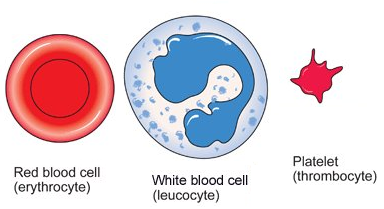
Blood cells - structure and functions - Biology Notes for IGCSE 2014
The structure of the heart - Structure and function of the ... It is located in the middle of the chest and slightly towards the left. The heart is a large muscular pump and is divided into two halves - the right-hand side and the left-hand side. The...
Structure and Function of the Heart
Label Internal Anatomy of The Heart Diagram | Quizlet Start studying Label Internal Anatomy of The Heart. Learn vocabulary, terms, and more with flashcards, games, and other study tools.
label any six of the parts of the heart a g in the below figure - Science - TopperLearning.com ...
Heart Labeling Quiz: How Much You Know About Heart Labeling? Here is a Heart labeling quiz for you. The human heart is a vital organ for every human. The more healthy your heart is, the longer the chances you have of surviving, so you better take care of it. Take the following quiz to know how much you know about your heart. Questions and Answers 1. What is #1? 2. What is #2? 3. What is #3? 4. What is #4?
31 Human Heart To Label - Labels Design Ideas 2020
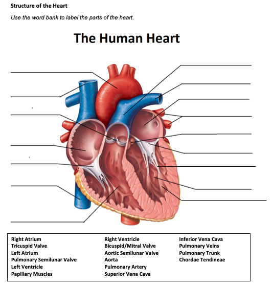
Heart Diagram with Labels and Detailed Explanation There are four chambers of the heart. The upper two chambers are the auricles and the lower two are called ventricles. There are four main valves of the human heart- aortic valve, mitral valve, pulmonary valve and tricuspid valve. They help prevent backflow of the blood.
labelled diagram of heart a level - Clip Art Library
Structure of the Heart | Biology for Majors II The heart is composed of three layers; the epicardium, the myocardium, and the endocardium, illustrated in Figure 1. The inner wall of the heart has a lining called the endocardium.The myocardium consists of the heart muscle cells that make up the middle layer and the bulk of the heart wall. The outer layer of cells is called the epicardium, of which the second layer is a membranous layered ...

The brain - structure and function - Cancer Information - Macmillan Cancer Support
The Heart - Science Quiz - Seterra The Heart - Science Quiz Home >> Seterra Anatomy and Science Quizzes >> The Heart The Heart - Science Quiz Aorta, Aortic valve, Left atrium, Left ventricle, Mitral valve, Pulmonary artery, Pulmonary valve, Pulmonary vein, Right atrium, Right ventricle, Septum, Superior vena cava, Tricuspid valve (13) Create custom quiz

Leadership: Group Work Can Be As Successful As You Want It To Be
Human Heart - Diagram and Anatomy of the Heart - Innerbody The heart is a muscular organ about the size of a closed fist that functions as the body's circulatory pump. It takes in deoxygenated blood through the veins and delivers it to the lungs for oxygenation before pumping it into the various arteries (which provide oxygen and nutrients to body tissues by transporting the blood throughout the body).

The chest x-ray in cardiovascular disease - wikidoc
Heart Anatomy: Labeled Diagram, Structures, Blood Flow ... Feb 24, 2021 · Chambers of the Heart Let's begin with the chambers of the heart. There are 4 chambers, labeled 1-4 on the diagram below. To help simplify things, we can convert the heart into a square. We will then divide that square into 4 different boxes which will represent the 4 chambers of the heart.
The Heart - Labelled diagram
Heart Diagram with Labels and Detailed Explanation - Byju's Diagram of Heart. The human heart is the most crucial organ of the human body. It pumps blood from the heart to different parts of the body and back to the heart. The most common heart attack symptoms or warning signs are chest pain, breathlessness, nausea, sweating etc. The diagram of heart is beneficial for Class 10 and 12 and is frequently ...

Katherina Krafts: Puffy 3D Origami Hearts
Label the heart — Science Learning Hub In this interactive, you can label parts of the human heart. Drag and drop the text labels onto the boxes next to the diagram. Selecting or hovering over a box will highlight each area in the diagram. Pulmonary vein Right atrium Semilunar valve Left ventricle Vena cava Right ventricle Pulmonary artery Aorta Left atrium Download Exercise Tweet

HR & BP - bright's blogs
Heart Anatomy Labeling Game This is an online quiz called Heart Anatomy Labeling Game There is a printable worksheet available for download here so you can take the quiz with pen and paper. Your Skills & Rank Total Points 0 Get started! Today's Rank -- 0 Today 's Points One of us! Game Points 19 You need to get 100% to score the 19 points available Actions

Fig 2 Gross Anatomy of the Heart (c)
The Anatomy of the Heart - Quiz 1 - Free Anatomy Quiz The heart - an image of the heart with blank labels attached The circulatory system - upper body image, with blank labels attached The circulatory system - lower body image, with blank labels attached The circulatory system - a PDF file of the upper and lower body for printing out to use off-line Articles :

QuickStudy | Nervous System Laminated Study Guide | Nervous system, Heart attack symptoms, Anatomy
Heart Anatomy: Heart Dissection The picture below shows an anterior view of the heart with the pericardium removed. The letters indicated in the text refer to the labels on the picture. The anterior surface of the heart is characterized by the presence of the large arteries leaving the base of the heart, the pulmonary trunk (H) and the aorta (G). The pulmonary trunk is the ...

William H. Peck - PAPYRUS OF NES MIN THE PAPYRUS OF NES-MIN: AN EGYPTIAN BOOK OF THE DEADDetroit ...
Label the Heart Quiz - PurposeGames.com Ummmmmmm . . . it's pretty self explanatory . . . you label the heart. Just remember one thing - you're looking at the heart like it's in someone else so right and left are switched around. This quiz has tags. Click on the tags below to find other quizzes on the same subject. Anatomy.

Hammond Biology- Structure of the Heart - Revision Notes in GCSE Biology
heart | Structure, Function, Diagram, Anatomy, & Facts The heart cavity is divided down the middle into a right and a left heart, which in turn are subdivided into two chambers. The upper chamber is called an atrium ...
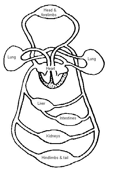
The Anatomy and Physiology of Animals/Circulatory System Worksheet - WikiEducator
Structure of the Heart - SEER Training The human heart is a four-chambered muscular organ, shaped and sized roughly like a man's closed fist with two-thirds of the mass to the left of midline. The heart is enclosed in a pericardial sac that is lined with the parietal layers of a serous membrane. The visceral layer of the serous membrane forms the epicardium. Layers of the Heart Wall
Post a Comment for "45 structure of the heart with labels"