44 cell diagram and labels
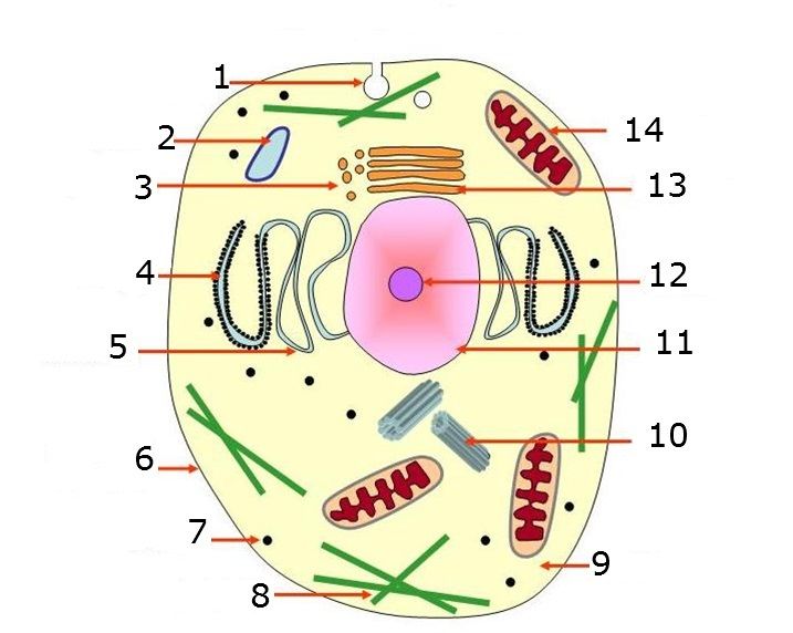
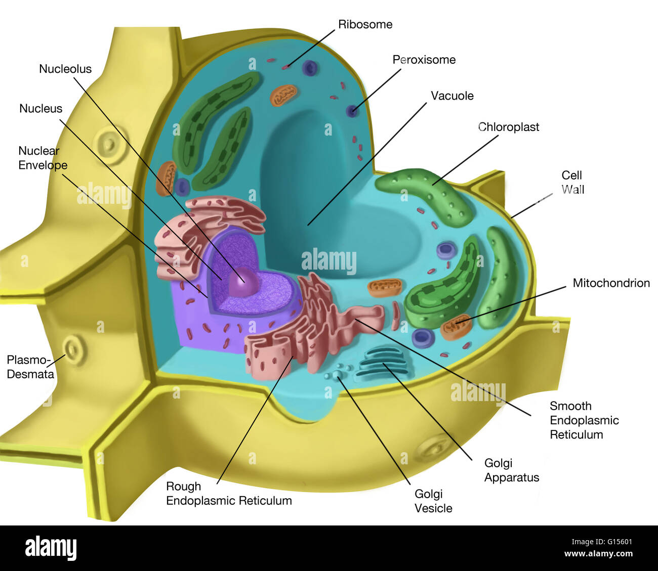
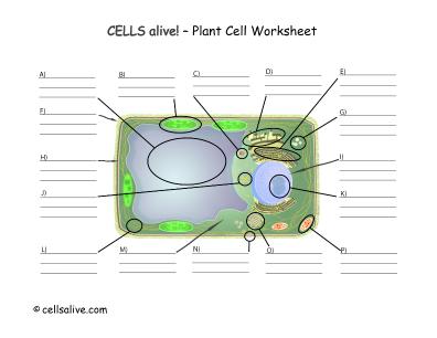
Plant Cell- Definition, Structure, Parts, Functions, Labeled Diagram Figure: Labeled diagram of plant cell, created with biorender.com. The typical characteristics that define the plant cell include cellulose, hemicellulose and pectin, plastids which play a major role in photosynthesis and storage of starch, large vacuoles responsible for regulating the cell turgor pressure. How to Add Labels to Scatterplot Points in Excel - Statology Step 3: Add Labels to Points. Next, click anywhere on the chart until a green plus (+) sign appears in the top right corner. Then click Data Labels, then click More Options…. In the Format Data Labels window that appears on the right of the screen, uncheck the box next to Y Value and check the box next to Value From Cells.
Interphase- Definition, Stages, Cell cycle, Diagram, Video The events of the cell cycle involve cell growth and cell division, of which the interphase defines the phase of cell growth where several metabolic reactions take place. The interphase is the preparation phase for mitosis and it is also the longest phase in the cell cycle. The interphase takes place in the cytoplasm and the cell nucleus.

Cell diagram and labels
consider this animal cell which organelle is labeled a The organelles in an animal cell are labeled. Which plant-cell organelle supports and maintains the cells shape and protects the cell from damage. A mitochondrion B Ribosome C Golgi Apparatus D Lysosome. Which organelle is labeled A. A cell is the basic unit of structure and function of all organisms. Drawing Of A Plant Cell With Labels : Animal Cell Diagram High ... Plant and animal cell organelles plus labeling them on a drawing. Browse draw and label plant cell resources on teachers pay teachers, a marketplace trusted by millions of teachers for original . Have been able to clearly see the parts of the cell and accurately label each part. Plant cell diagram science cells, plant science, science biology,. Alveoli: Anatomy, function and clinical points | Kenhub Membranes have a total thickness of only 0.5-micrometers, in contrast to the 7.5-micrometer diameter of the erythrocytes (blood cells) that pass through the capillaries. Want to keep learning about the anatomy of the respiratory system? Try our respiratory system quizzes and labeled diagrams.
Cell diagram and labels. Simple Labeled Cell Membrane Structure - Cellmembrane The cell wall also bonds with other cell walls to form the structure of the plant. Cell wall a thick, rigid membrane that surrounds a plant cell. Labeled diagram simple cuboidal epithelial cells are shaped like cubes, and the nucleus of each cell is large and located close to the center of the cell. Dec 13, 2021 · simple cuboidal epithelium: Cellular Respiration Equation, Types, Stages, Products & Diagrams Cellular Respiration Equation: Every machine needs specific parts and fuel in order to function. Likewise, "biological machines" also require well engineered parts and good energy source in order to work.Perhaps the second most important molecule (DNA is the first) is adenosine triphosphate (also known as ATP).Basically, ATP serves as the main energy currency of the cell. › innovation › galvanic-cell-workGalvanic Cell: Definition, Diagram and Working - Science ABC Jan 17, 2022 · Galvanic Cell vs Electrolytic Cell. Lastly, once dead, galvanic cells cannot be revived or recharged. This is why one must change the batteries in an alarm clock or remote control from time to time. The kind of electrochemical cell that can be recharged is an electrolytic cell. byjus.com › biology › liver-diagramLiver Diagram with Detailed Illustrations and Clear Labels Liver Diagram The liver is one of the most important organs in the human body. Anatomically, the liver is a meaty organ that consists of two large sections called the right and the left lobe.
Labeling the Animal Cell Quiz - purposegames.com This is an online quiz called Labeling the Animal Cell. There is a printable worksheet available for download here so you can take the quiz with pen and paper. Your Skills & Rank. Total Points. 0. Get started! Today's Rank--0. Today 's Points. One of us! Game Points. 9. You need to get 100% to score the 9 points available. Plant and Animal Cell Worksheets and Animal Cell Project Grade 6 There is a blank animal cell diagram and a blank plant cell diagram in both black and white and color options. ... Suggested Grades 4-6. She features awesome colorful printables that label both the animal and plant cells, notebook pages, blank forms for your student to use to label the cells themselves, coloring pages, Venn diagram and word ... Simple Animal Cell Drawing With Labels / Draw A Labelled Diagram Of The ... 5th grade science and biology. Animal cells differ from plant . Lower, middle and higher ability versions are available. Draw A Labelled Diagram Of The Animal Cell Sarthaks Econnect Largest Online Education Community from Printable animal cell diagram to help you learn the organelles in an animal cell in preparation for your test or quiz. Animal Cells: Labelled Diagram, Definitions, and Structure Cilia and Flagella. Some eukaryotic cells either have cilia or flagella. Cilia are small, wiggling arm-like structures, whereas flagella are like a tail. Both structures are made of long protein fibers called microtubules, with a structure where nine microtubules form a ring around two central microtubules.
Structure Animal Cell Easy Drawing And Label / 3 950 Human Cell Diagram ... Printable Animal Cell Diagram Labeled Unlabeled And Blank from timvandevall.com In this printable, students broaden their horizons to know the structure of an atom that includes the nucleus, protons, neutrons, electrons, and electron shells. Study each new biological concept and process thoroughly before moving on to the next level. Animal Cell Label The Organelles In The Diagram Below : 1 2 Difference ... To get an answer to your question ️ label: Animal cell organelles and functions with diagrams. Label The Following For The Bacterial Cells Animal Chegg Com from media.cheggcdn.com Using the letters from the table in model 1, label the cell diagram with the organelle names. Label the organelles in the diagram below of a typical plant cel. Plant and Animal Cell: Labeled Diagram, Structure, Function - Embibe The variation in cell content between plants and animals, as well as their structure and functions, is the source of these differences. Each sensory organ in a cell has a specific job to do. Some cell organelles are found in both plant and animal cells, whereas others are specific to one or the other. Animal Cell Labeling Quiz Questions And Answers - ProProfs Take this animal cell labeling quiz to learn more about this topic! Animal cell labeling can be tricky at first, so why don't we start you off relatively easy with this animal cell part labeling quiz! In this one, we'll be giving you a question referring to a given diagram and asking you to label it. Simple, right? Let's find out how many you get right. All the best! Let's go!
[DIAGRAM] Cell Diagram Labeling
Cell Organelles- Definition, Structure, Functions, Diagram An additional non-living layer present outside the cell membrane in some cells that provides structure, protection, and filtering mechanism to the cell is the cell wall. Structure of Cell Wall. In a plant cell, the cell wall is made up of cellulose, hemicellulose, and proteins while in a fungal cell, it is composed of chitin. A cell wall is ...

Inspired Class: Create 3D Animal Cells with Play Doh
› cells › bactcellInteractive Bacteria Cell Model - CELLS alive In the space are enzymes and other proteins that help digest and move nutrients into the cell. Cell Wall: Composed of peptidoglycan (polysaccharides + protein), the cell wall maintains the overall shape of a bacterial cell. The three primary shapes in bacteria are coccus (spherical), bacillus (rod-shaped) and spirillum (spiral).
Animal Cell No Label - Plant Cell Diagram Without Labels Plant Cell Diagram Plant Cells ...
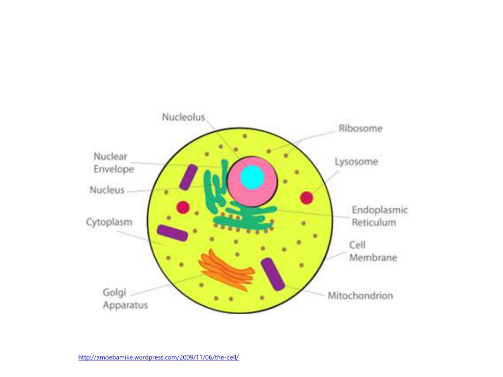
Learn the parts of a cell with diagrams and cell quizzes Labeled cell diagram. For this exercise we'll start with an image of a cell diagram ready labeled. Study this and make sure that you're clear about which structure is found where. Cell diagram unlabeled. It's time to label the cell yourself! As you fill in the cell structure worksheet, remember the functions of each part of the cell that ...
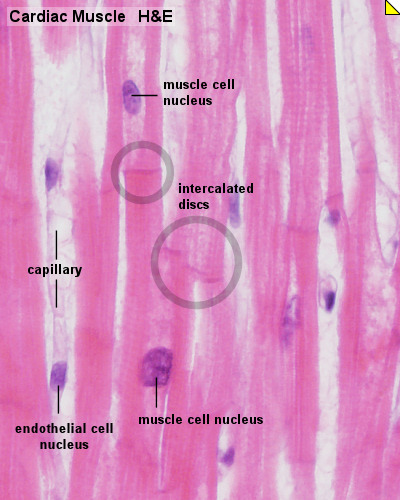
Cardiac Muscle Histology - Embryology
Eukaryotic Cell Drawing And Labeling - 12 images - eukaryotic cell ... Here are a number of highest rated Eukaryotic Cell Drawing And Labeling pictures on internet. We identified it from obedient source. Its submitted by dealing out in the best field. We say you will this nice of Eukaryotic Cell Drawing And Labeling graphic could possibly be the most trending topic considering we allocation it in google help or ...
CELL - Labelled diagram
Draw A Labelled Diagram Of Animal Cell And Plant Cell - DRAW IT NEAT ... See how a generalized structure of an animal cell and plant cell look with labeled diagrams. The largest organelle within the cell. 85kb, plant and animal cell diagram labeled drawing picture with tags: Asked nov 28, 2017 in class ix science by ashu premium (930 points). A system of flattened membranes called cisternae (mainpoint:

Sel Mast (Mast Cell)
Electrochemistry: Galvanic Cells and the Nernst Equation The following is an electrochemical cell diagram for the reaction shown in the movie above: Zn(s) + Cu 2+ ... + Cu(s). Match the following labels to their location on the diagram. You will not use all of the labels. Oxidation half-cell. Reduction half-cell. Direction of anion flow in salt bridge >>> Direction of anion flow in salt bridge ...
Plant cell structure. Artwork of a sectioned plant cell. The features Stock Photo - Alamy
Drag the correct labels to the image. Not all labels will be used ... The cell organelles that Ricky is likely to associate with the parts of the egg would be plasma membrane, cytoplasm, and nucleus respectively.. Cell Organelles. Cell organelles refer to the component parts of a cell with specific functions.. From the image, the fried egg of Ricky looks like the structure of a typical animal cell.. The outermost part of an animal cell that delimits the entire ...
Plant Cell Diagram With Labels And Functions - Diagram Media
Cell Membrane (Plasma Membrane) - Genome.gov The cell membrane, also called the plasma membrane, is found in all cells and separates the interior of the cell from the outside environment. The cell membrane consists of a lipid bilayer that is semipermeable. The cell membrane regulates the transport of materials entering and exiting the cell. If playback doesn't begin shortly, try ...

ROOT HAIR CELL CAKE | My son's biology homework was to make … | Flickr
chemostratigraphy.com › how-to-plot-a-ternaryHow to plot a ternary diagram in Excel Feb 13, 2022 · Adding labels to the apices. Next, we need some space for the apices labels: click into the Plot Area (not the Chart Area) then resize by holding the Shift key (this ensures an equal scaling) and use the mouse cursor on one of the corner pick-points. Then recentre the Plot Area in the Chart Area.
Blank Animal Cell Diagram To Label Pdf - Diagram Media
› cell_cycle_jsInteractive Cell Cycle - CELLS alive INTERPHASE. Gap 0. Gap 1. S Phase. Gap 2. MITOSIS . ^ Cell Cycle Overview Cell Cycle Mitosis > Meiosis > Get the Cell Division PowerPoints

circulatory Diagram Of Cardiovascular System system diagrams are visual representations of the ...
byjus.com › biology › skin-diagramSkin Diagram with Detailed Illustrations and Clear Labels Skin Diagram The largest organ in the human body is the skin, covering a total area of about 1.8 square meters. The skin is tasked with protecting our body from the external elements as well as microbes.

Alila Medical Media | Medical Genetics Images & Videos
Plant Cells: Labelled Diagram, Definitions, and Structure Plastids and Chloroplasts. Plants make their own food through photosynthesis. Plant cells have plastids, which animal cells don't. Plastids are organelles used to make and store needed compounds. Chloroplasts are the most important of plastids. They convert light energy from the sun into sugar and oxygen. The most exposed parts of the plants ...

draw the diagrams of different types of cells and label them - Brainly.in
Male Reproductive System: Labeled Diagram of Organs - Video & Lesson ... Learn the structure and functions of male reproductive anatomy with these labeled diagrams. Updated: 07/14/2021 Table of Contents ... Both reproductive systems are vital to produce sex cells ...

A&PII Eportfolio: Objective 54: How erythropoietin regulates red blood cell,production
Structure of Cell: Definition, Types, Diagram, Functions - Embibe A cell structure is composed of many components which are present inside the cell. These components carry out the various important functions which are important in the main functioning of the cell. Various kinds of cells show special differences, yet they all have some basic structural plan consisting of three essential parts: (i) cell ...

Brown Adipocytes
Cells Diagram | Science Illustration Solutions - Edrawsoft Cells Diagram. Cells are the basic building blocks of all living things. The human body is composed of trillions of cells. Cells have many parts, each with a different function. Some of these parts, called organelles, are specialized structures that perform certain tasks within the cell. Drawing cells diagram helps you better understand your ...
Chapter 3 Review
Diagram of Human Heart and Blood Circulation in It Four Chambers of the Heart and Blood Circulation. The shape of the human heart is like an upside-down pear, weighing between 7-15 ounces, and is little larger than the size of the fist. It is located between the lungs, in the middle of the chest, behind and slightly to the left of the breast bone. The heart, one of the most significant organs ...
Post a Comment for "44 cell diagram and labels"